51
ENFERMEDAD ÁCIDO PÉPTICA Y ERGE. N. Angélica Rebollar Martínez

ENFERMEDAD ÁCIDO PÉPTICA Y ERGE.
N. Angélica Rebollar Martínez
DEFINICIÓN
Villalobos P, Olivera M.A. “Gastroenterología”. 5ª edición. Méndez Editores. Pag. 183-193.

Epidemiología
Prevalencia del 5-10% Multifactorial Úlcera gástrica (frecuente – 64%) Úlcera duodenal 30% Uso crónico de AINES (40%). Presencia de H. pylori (50%).
Villalobos P, Olivera M.A. “Gastroenterología”. 5ª edición. Méndez Editores. Pag. 183-193. c

Villalobos P, Olivera M.A. “Gastroenterología”. 5ª edición. Méndez Editores. Pag. 183-193.
Fisiopatología
Multifactorial Mucosa y HCO3 H. pylori gastritis antral no atrófica
aumento de la secreción. - Producción de ureasa - Variación de glucoproteínas en moco. - Aumento de la permeabilidad, lipasa
y fosfolipasa.
Villalobos P, Olivera M.A. “Gastroenterología”. 5ª edición. Méndez Editores. Pag. 183-193.

Manifestaciones clínicas.
Dolor en epigastrio - Sensación de ardor - Ritmo es pospandrial tardío Hiporexia Malestar general Hemorragia (1er síntoma). Nausea y vomito. Pirosis /regurgitación Eructos
Villalobos P, Olivera M.A. “Gastroenterología”. 5ª edición. Méndez Editores. Pag. 183-193.

Villalobos P, Olivera M.A. “Gastroenterología”. 5ª edición. Méndez Editores. Pag. 183-193.
Helicobacter pylori.
Cag A - Proteína de la membrana externa - Inmunogénica - Adhieren a las células del epitelio
gástrico secreción de mediadores inflamatorios IL-8 NF-kB.
- Elevación de niveles IgG: Úlcera duodenal 87.5% Úlcera gástrica 76%
Cervantes G. Helicobacter pylori e infecciones asociadas. www.ejournal .unam.mx.

Prueba de ureasa
Hidroliza la urea por acción de la ureasa - Iones de amonio - Aumenta el pH cambio de coloración CLO-test Prueba de aliento con urea marcada 13C o
14C - Cápsulas - 15-30 minutos. PCR
Cervantes G. Helicobacter pylori e infecciones asociadas. www.ejournal .unam.mx.

Helicobacter pylori.
Importante el tx por las recidivas. 500mg claritromicina + 1 gr amoxicilina +
IBP (omeprazol 20 mg, lansoprazol 30mg, esomeprazol 40 mg) c/12 hrs durante 2 semanas.
Inhibidor de H2: ranitidina 300mg c/12 hrs x 2 sem y 300mg x la noche 1 mes.
Ranitidina + subsalicilato de bismuto 1 tab c/12 hrs x 1 mes.
80% cicatrización a 4-6 semanas. Villalobos P, Olivera M.A. “Gastroenterología”. 5ª edición. Méndez Editores. Pag. 183-193.

TRATAMIENTO
TRATAMIENTO QX.
Hemorragia persistente o grave Perforación Penetración Estenosis Úlcera rebelde a tratamiento médico.
Villalobos P, Olivera M.A. “Gastroenterología”. 5ª edición. Méndez Editores. Pag. 183-193.

ENFERMEDAD POR REFLUJO GASTROESOFÁGICO

DEFINICION
Nimish V et col. “ The Montreal Definition and Classification of Gastroesophageal Reflux Disease: A Global Evidence –Based Consensus”. Am
J Gastroenterol 2006;101:1900-1920.c
EPIDEMIOLOGÍA
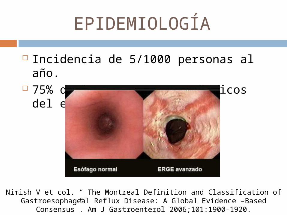
Incidencia de 5/1000 personas al año. 75% de los procesos patológicos del
esófago.
Nimish V et col. “ The Montreal Definition and Classification of Gastroesophageal Reflux Disease: A Global Evidence –Based Consensus”. Am
J Gastroenterol 2006;101:1900-1920.c
FISIOPATOLOGÍA

Richter Joel et col. “The Many Manifestations of Gastroesophageal Reflux, Disease: Presentastion, Evaluation, and Treatment. Gastroenterol Clin N Am.
2007:36;577-599.

Richter Joel et col. “The Many Manifestations of Gastroesophageal Reflux, Disease: Presentastion, Evaluation, and Treatment. Gastroenterol Clin N Am.
2007:36;577-599.

Richter Joel et col. “The Many Manifestations of Gastroesophageal Reflux, Disease: Presentastion, Evaluation, and Treatment. Gastroenterol Clin N Am.
2007:36;577-599.

Richter Joel et col. “The Many Manifestations of Gastroesophageal Reflux, Disease: Presentastion, Evaluation, and Treatment. Gastroenterol Clin N Am.
2007:36;577-599.

Richter Joel et col. “The Many Manifestations of Gastroesophageal Reflux, Disease: Presentastion, Evaluation, and Treatment. Gastroenterol Clin N Am.
2007:36;577-599.

FACTORES DE RIESGO
Edad Sexo (masculino). Sobrepeso y Obesidad (IMC >25). Embarazo (48-79%). Alcoholismo Ejercicio
Richter Joel et col. “The Many Manifestations of Gastroesophageal Reflux, Disease: Presentastion, Evaluation, and Treatment. Gastroenterol Clin N Am.
2007:36;577-599.
FACTORES DE RIESGO
Infección por Helicobacter pylori. Genético: cromosoma 13 (pediátricos). Hernia hiatal, alteraciones en la
motilidad esofágica. Ingesta de medicamentos: teofilina,
nitratos, anticonceptivos orales, agonista B2 adrenérgicos.
Richter Joel et col. “The Many Manifestations of Gastroesophageal Reflux, Disease: Presentastion, Evaluation, and Treatment. Gastroenterol Clin N Am.
2007:36;577-599.

MANIFESTACIONES CLÍNICAS
ESOFÁGICAS Pirosis (garganta/región interescapular). Regurgitación Disfagia Hematemesis /melena (2-6%) Hipo Halitosis Eructos Sialorrea Tos
Richter Joel et col. “The Many Manifestations of Gastroesophageal Reflux, Disease: Presentastion, Evaluation, and Treatment. Gastroenterol Clin N Am.
2007:36;577-599.

MANIFESTACIONES CLÍNICAS
EXTRAESOFÁGICAS Dolor torácico Disfonía Dolor faríngeo Otitis Laringitis Pérdida del esmalte de los dientes. Asma bronquial Neumonía Apnea Bronquiectasias
Richter Joel et col. “The Many Manifestations of Gastroesophageal Reflux, Disease: Presentastion, Evaluation, and Treatment. Gastroenterol Clin N Am.
2007:36;577-599.

Richter Joel et col. “The Many Manifestations of Gastroesophageal Reflux, Disease: Presentastion, Evaluation, and Treatment. Gastroenterol Clin N Am.
2007:36;577-599.

DIAGNÓSTICO
pH-metría esofágica - Reflujo gastroesofágico patológico - Cuantificar el número de episodios - Si ocurre de pie o decúbito - % de tiempo en el que el pH < 4. - Relación de síntomas con los episodios. - Índice DeMeester positivo > 14. Sensibilidad 78%
Kenneth R, et col. “ Updated Guidelines for the Diagnosis and Treatment of Gastroesophageal Reflux Disease. Am J Gastroenterol 2005; 100:190-200.

pHmetría esofágica.
Suárez Moran E, et col. Frecuencia de las diferentes variantes de la enfermedad por reflujo gastroesofágico no erosiva. Rev Med Hosp Gen
Mex 2006; 69:12-16
DIAGNÓSTICO
MANOMETRIA ESOFÁGICA. Prueba indicada para la conocer la
función mecánica del EEI. Peristalsis esofágica. Tratamiento quirúrgico.
Kenneth R, et col. “ Updated Guidelines for the Diagnosis and Treatment of Gastroesophageal Reflux Disease. Am J Gastroenterol 2005; 100:190-200.

DIAGNÓSTICO
IMPEDANCIA ELÉCTRICA INTRALUMINAL (IEI)
Detectar flujo retrógrado de contenido gástrico hacia el esófago.
Independiente del pH. 6 sitios: 4 distales y 2 proximales . - 3,5,7,9,15 y 17 cm por arriba del EEI. Sensor del pH se encuentra en el 2º
segmento (5 cm por arriba EEI). Kenneth R, et col. “ Updated Guidelines for the Diagnosis and Treatment of Gastroesophageal Reflux Disease. Am J Gastroenterol 2005; 100:190-200.
DIAGNÓSTICO
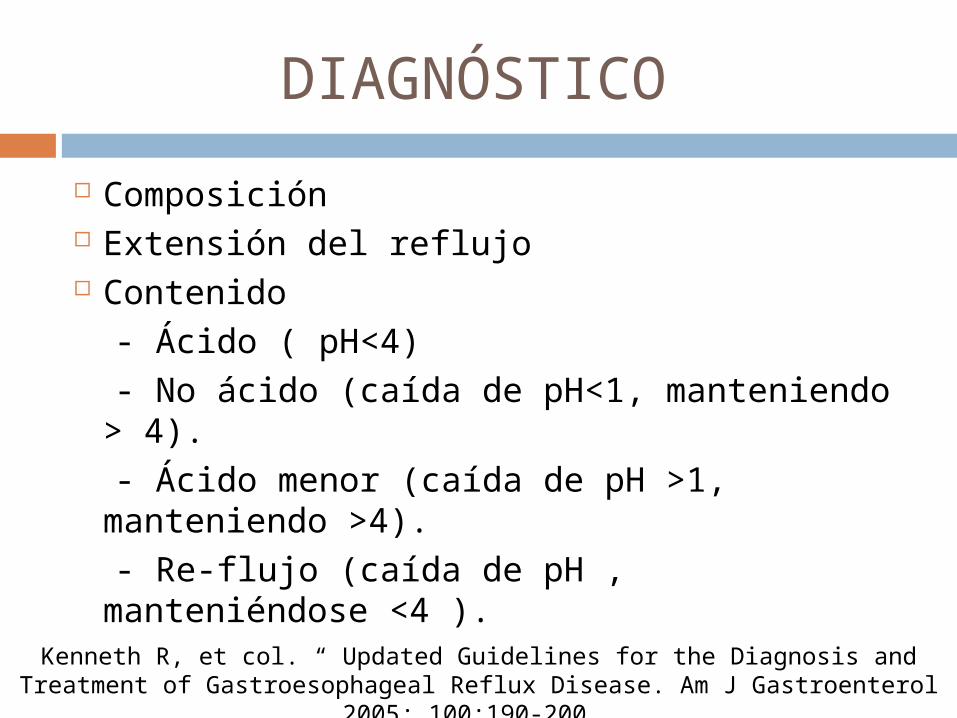
Composición Extensión del reflujo Contenido - Ácido ( pH<4) - No ácido (caída de pH<1, manteniendo >
4). - Ácido menor (caída de pH >1, manteniendo
>4). - Re-flujo (caída de pH , manteniéndose <4 ).
Kenneth R, et col. “ Updated Guidelines for the Diagnosis and Treatment of Gastroesophageal Reflux Disease. Am J Gastroenterol 2005; 100:190-200.

DIAGNÓSTICO
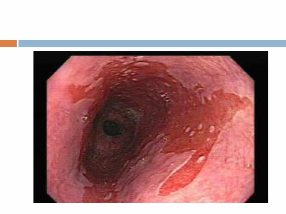
Endoscopía - Esofagitis - Sensibilidad 50% - Especificidad 90-95%. - Biopsia - Complicaciones: EB
Kenneth R, et col. “ Updated Guidelines for the Diagnosis and Treatment of Gastroesophageal Reflux Disease. Am J Gastroenterol 2005; 100:190-200.

CLASIFICACIÓN DE ESOFAGITIS (LA)
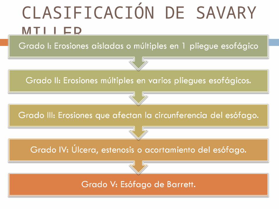
CLASIFICACIÓN DE SAVARY MILLER
DIAGNÓTICO
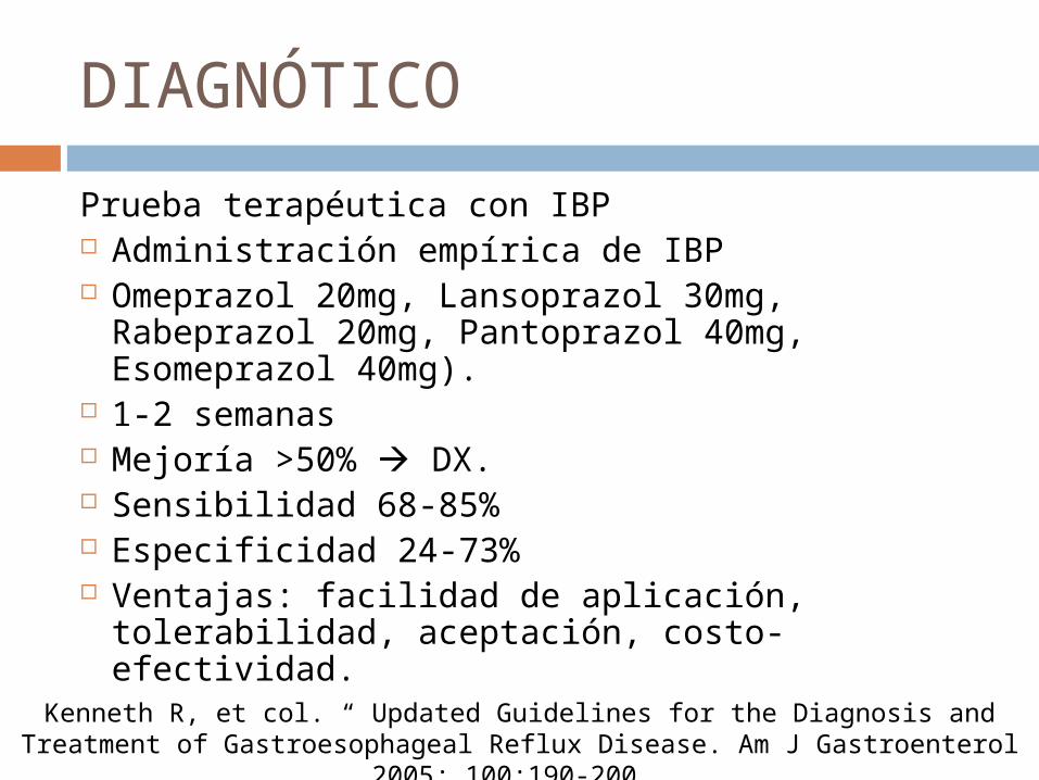
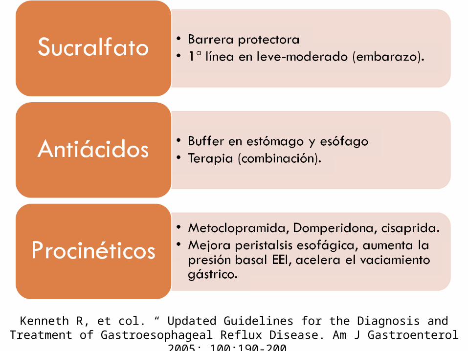
Prueba terapéutica con IBP Administración empírica de IBP Omeprazol 20mg, Lansoprazol 30mg,
Rabeprazol 20mg, Pantoprazol 40mg, Esomeprazol 40mg).
1-2 semanas Mejoría >50% DX. Sensibilidad 68-85% Especificidad 24-73% Ventajas: facilidad de aplicación,
tolerabilidad, aceptación, costo-efectividad. Kenneth R, et col. “ Updated Guidelines for the Diagnosis and Treatment of Gastroesophageal Reflux Disease. Am J Gastroenterol 2005; 100:190-200.
Kenneth R, et col. “ Updated Guidelines for the Diagnosis and Treatment of Gastroesophageal Reflux Disease. Am J Gastroenterol 2005; 100:190-200.
Kenneth R, et col. “ Updated Guidelines for the Diagnosis and Treatment of Gastroesophageal Reflux Disease. Am J Gastroenterol 2005; 100:190-200.

Kenneth R, et col. “ Updated Guidelines for the Diagnosis and Treatment of Gastroesophageal Reflux Disease. Am J Gastroenterol 2005; 100:190-200.

TRATAMIENTO
QUIRÚRGICO 20-50 % tienen ERGE persistente
progresivo. Pacientes que dependen del Tx. IBP o que
necesitan dosis cada vez mayores para control de la enfermedad.
Detección esofagitis grave (endoscopia) + deficiencia estructural EEI.
Estenosis a pesar de Tx médico. Biopsia: displasia grave o carcinoma
intramucoso. The Medical Clinics of North America. “Clinical presentation, diagnosis, and management of gastroesophageal reflux disease. Med Clin N Am 89;2005:
243-291.
TRATAMIENTO
QUIRÚRGICO El principal objetivo es restaurar de
manera segura la estructura del esfínter o evitar su acortamiento durante la distensión gástrica, en tanto se conserva la capacidad del paciente para deglutir con normalidad.
5 principios para la reconstrucción del cardias.
Schwartz. “Principios de cirugía”. 8ª edición. Vol I. Ed. Mc Graw Hill. Pag. 835- 873.

Schwartz. “Principios de cirugía”. 8ª edición. Vol I. Ed. Mc Graw Hill. Pag. 835- 873.

Schwartz. “Principios de cirugía”. 8ª edición. Vol I. Ed. Mc Graw Hill. Pag. 835- 873.
TRATAMIENTO
Funduplicatura tipo Nissen - Laparoscopía F. tipo Nissen transtorácica. F. parciales laparoscópicas : Toupet y
Guarner. - 270º 90% alivio de síntomas. Mortalidad 10-15%
Schwartz. “Principios de cirugía”. 8ª edición. Vol I. Ed. Mc Graw Hill. Pag. 835- 873.


COMPLICACIONES
Estenosis péptica - Dilataciones esofágicas periódicas. - Control adecuado del reflujo.
Esofagitis hemorrágica. - Hemorragias agudas (7%) - Esófago distal
Nimish V et col. “ The Montreal Definition and Classification of Gastroesophageal Reflux Disease: A Global Evidence –Based Consensus”. Am
J Gastroenterol 2006;101:1900-1920.c
COMPLICACIONES
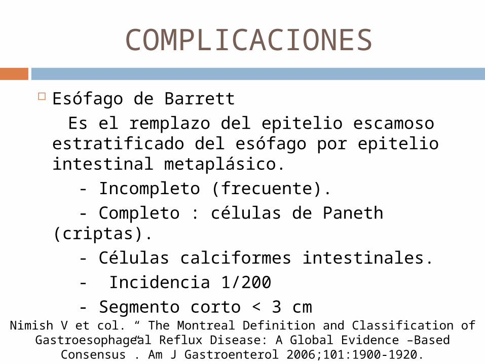
Esófago de Barrett Es el remplazo del epitelio escamoso
estratificado del esófago por epitelio intestinal metaplásico.
- Incompleto (frecuente). - Completo : células de Paneth (criptas). - Células calciformes intestinales. - Incidencia 1/200 - Segmento corto < 3 cmNimish V et col. “ The Montreal Definition and Classification of
Gastroesophageal Reflux Disease: A Global Evidence –Based Consensus”. Am J Gastroenterol 2006;101:1900-1920.
c

Células de Paneth

COMPLICACIONES
Esófago de Barrett - Lesión premaligna Adenocarcinoma
(10%) - Multifactorial Biopsia Displasia - Bajo grado : difícil dx. - Alto grado
Nimish V et col. “ The Montreal Definition and Classification of Gastroesophageal Reflux Disease: A Global Evidence –Based Consensus”. Am
J Gastroenterol 2006;101:1900-1920.c
BIBLIOGRAFÍA
Schwartz. “Principios de cirugía”. 8ª edición. Vol I. Ed. Mc Graw Hill. Pag. 835- 873.
Villalobos P, Olivera M.A. “Gastroenterología”. 5ª edición. Méndez Editores. Pag. 183-193.
Richter Joel et col. “The Many Manifestations of Gastroesophageal Reflux, Disease: Presentastion, Evaluation, and Treatment. Gastroenterol Clin N Am. 2007:36;577-599.
Nimish V et col. “ The Montreal Definition and Classification of Gastroesophageal Reflux Disease: A Global Evidence –Based Consensus”. Am J Gastroenterol 2006;101:1900-1920.
Kenneth R, et col. “ Updated Guidelines for the Diagnosis and Treatment of Gastroesophageal Reflux Disease. Am J Gastroenterol 2005; 100:190-200.
Catarci M. Gentileschi P. “Evidence-based Appraisal of Antireflux Fundoplication”. Ann Surg 2004; 239: 325-337.
The Medical Clinics of North America. “Clinical presentation, diagnosis, and management of gastroesophageal reflux disease. Med Clin N Am 89;2005: 243-291.


![Vomitos ERGE 2012[1]](https://static.documentos.tech/doc/80x56/55721058497959fc0b8d074d/vomitos-erge-20121.jpg)


