BIM and mTOR expression levels predict outcome to erlotinib in EGFR-mutant non-small-cell lung cancer Niki Karachaliou 1 , Jordi Codony-Servat 2 , Cristina Teixido 2 , Sara Pilotto 3 , Ana Drozdowskyj 4 , Carles Codony-Servat 2 , Ana Gimenez-Capitan 2 , Miguel Angel Molina-Vila 2 , Jordi Bertran-Alamillo 2 , Radj Gervais 5 , Bartomeu Massuti 6 , Teresa Moran 7 , Margarita Majem 8 , Enriqueta Felip 9 , Enric Carcereny 7 , Rosario Garcia- Campelo 10 , Santiago Viteri 1 , Maria Gonzalez Cao 1 , Daniela Morales-Espinosa 1 , Alberto Verlicchi 11 , Elisabetta Crisetti 12 , Imane Chaib 7 , Mariacarmela Santarpia 13 Jose Luis Ramirez 7 , Joaquim Bosch-Barrera 14 , Andres Felipe Cardona 15 , Filippo de Marinis 16 , Guillermo Lopez Vivanco 17 , Jose Miguel Sanchez 18 , Alain Vergnenegre 19 , Jose Javier Sanchez Hernandez 20 , Isabella Sperduti 21 , Emilio Bria 3 , *Rafael Rosell 1,2,7,22,23 1 Instituto Oncológico Dr Rosell, Quiron-Dexeus University Hospital, Barcelona, Spain; 2 Pangaea Biotech, Barcelona, Spain; 3 Department of Medical Oncology, University of Verona, Azienda Ospedaliera Universitaria Integrata, Verona, Italy; 4 Pivotal, Madrid, Spain; 5 Centre François Baclesse, Caen, France; 6 Hospital General de Alicante, Alicante, Spain; 7 Catalan Institute of Oncology, Hospital Germans Trias i Pujol, Badalona, Spain; 8 Hospital de Sant Pau, Barcelona; 9 Hospital Vall d’Hebron, Barcelona, Spain; 10 Complexo Hospitalario Universitario La Coruña, Spain; 11 Ospedale Santa Maria delle Croci, Ravenna, Italy; 12 Department of Medical and Surgical Sciences, Institute of Respiratory Diseases, University of Foggia, Italy; 13 Medical Oncology Unit, Human Pathology Department, University of Messina, Italy; 14 Catalan Institute of Oncology, Hospital Josep Trueta, Girona, Spain, 15 Clinical and Traslational Oncology Group, Institute of Oncology, Clínica del Country, Bogotá, Colombia; 16 Direttore, Divisione di Oncologica Toracica, Istituto Europeo di Oncologia – IEO, Milano, Italy; 17 Chief, Medical Oncology Service, Hospital de Cruces, Barakaldo, Vizcaya, Spain; 18 Medical Oncology Service, Hospital de la Princesa, Madrid, Spain; 19 Hôpital du Cluzeau, Limoges, France; 20 Unidad de Investigación en Salud Pública CIDICS-UANL, 21 Biostatistics, Regina Elena National Cancer Institute, Rome, 22 Molecular Oncology Research (MORe) Foundation, Barcelona, Spain; 23 Germans Trias i Pujol Health Sciences Institute and Hospital, Campus Can Ruti
Transcript
BIM and mTOR expression levels predict outcome to erlotinib in EGFR-mutant
non-small-cell lung cancer
Niki Karachaliou1, Jordi Codony-Servat2, Cristina Teixido2, Sara Pilotto3, Ana
Drozdowskyj4, Carles Codony-Servat2, Ana Gimenez-Capitan2, Miguel Angel
Campelo10, Santiago Viteri1, Maria Gonzalez Cao1, Daniela Morales-Espinosa1,
Alberto Verlicchi11, Elisabetta Crisetti12, Imane Chaib7, Mariacarmela Santarpia13
Jose Luis Ramirez7, Joaquim Bosch-Barrera14, Andres Felipe Cardona15, Filippo de
Marinis16, Guillermo Lopez Vivanco17, Jose Miguel Sanchez18, Alain Vergnenegre19,
Jose Javier Sanchez Hernandez20, Isabella Sperduti21, Emilio Bria3, *Rafael
Rosell1,2,7,22,23
1Instituto Oncológico Dr Rosell, Quiron-Dexeus University Hospital, Barcelona, Spain; 2Pangaea Biotech, Barcelona, Spain; 3Department of Medical Oncology, University of Verona, Azienda Ospedaliera Universitaria Integrata, Verona, Italy; 4Pivotal, Madrid, Spain; 5Centre François Baclesse, Caen, France; 6Hospital General de Alicante, Alicante, Spain; 7Catalan Institute of Oncology, Hospital Germans Trias i Pujol, Badalona, Spain; 8 Hospital de Sant Pau, Barcelona; 9Hospital Vall d’Hebron, Barcelona, Spain; 10 Complexo Hospitalario Universitario La Coruña, Spain; 11 Ospedale Santa Maria delle Croci, Ravenna, Italy; 12 Department of Medical and Surgical Sciences, Institute of Respiratory Diseases, University of Foggia, Italy;
13Medical Oncology Unit, Human Pathology Department, University of Messina, Italy; 14Catalan Institute of Oncology, Hospital Josep Trueta, Girona, Spain, 15 Clinical and Traslational Oncology Group, Institute of Oncology, Clínica del Country, Bogotá, Colombia; 16Direttore, Divisione di Oncologica Toracica, Istituto Europeo di Oncologia – IEO, Milano, Italy; 17Chief, Medical Oncology Service, Hospital de Cruces, Barakaldo, Vizcaya, Spain; 18Medical Oncology Service, Hospital de la Princesa, Madrid, Spain; 19Hôpital du Cluzeau, Limoges, France; 20Unidad de Investigación en Salud Pública CIDICS-UANL, 21Biostatistics, Regina Elena National Cancer Institute, Rome, 22Molecular Oncology Research (MORe) Foundation, Barcelona, Spain; 23Germans Trias i Pujol Health Sciences Institute and Hospital, Campus Can Ruti
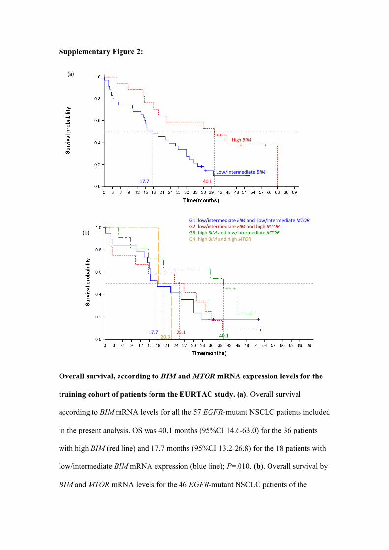
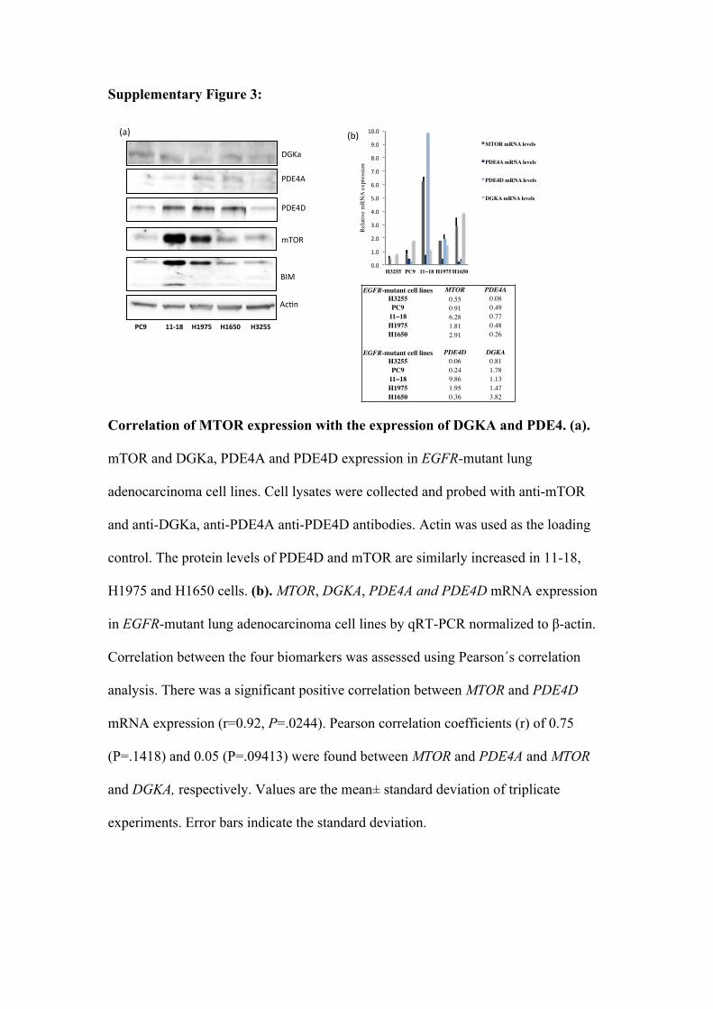
Supplementary Material
Supplementary Table 1. Patient characteristics of the 19 patients included in the
validation cohort
Clinical parameter N(%) Sex Female 14 (73.7) Male 5 (26.3) Age group <65 years 11 (77.9) >= 65 years 8 (42.1) Smoking status Never smoker 15 (78.9) Former smoker 3 (15.8) Current Smoker 1 (5.3) ECOG PS 0-1 12 (63.2) 2 7 (36.8) Histologic Diagnosis Adenocarcinoma 19 (100.0) Clinical Stage IIIA 1 (5.3) IIIB 2 (10.5) IV 16 (84.2) Bone metastasis Yes 9 (47.4) No 10 (52.6) Brain metastasis Yes 7 (36.8) No 12 (63.2) Other metastasis Yes 12 (63.2) No 7 (36.8) Type of EGFR mutation del19 12 (63.2) L858R 7 (36.8) Type of EGFR TKI Erlotinib 13 (68.4) Gefitinib 5 (26.3) Afatinib 1 (5.3) Response Complete response 2 (10.5) Partial response 10 (52.6) Stable disease 7 (36.8)
Supplementary Table 2: Univariate analyses of progression-free and overall survival
in 57 patients from the EURTAC trial included in the present study.
PFS OS Variable HR (95%CI) P HR (95%CI) P Gender Female 1.00 - 1.00 Male 1.14 (0.60, 2.18) .6859 1.20 (0.63, 2.28) .5773 Smoking history Current smoker 1.00 1.00 Never smoked 1.10 (0.33, 3.68) .8737 0.39 (0.15, 1.05) .1791 Former smoker 1.05 (0.30, 3.68) .9391 0.46 (0.17, 1.31 .1472 ECOG performance status